
Fig. 2.
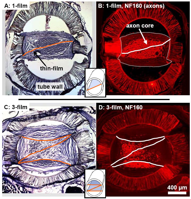
Representative cross-sections from a 1-film (A,B) and 3-film (C,D) channel at the 13 week time point. False colored lines mark the locations of the aligned thin-films. (A,C): Tissue stained cross-sections taken 3.5 mm into the nerve gap. The location and structure of the regenerated tissue cables were visibly influenced by the thin-films. (B,D): Cross-sections taken from 2.5 mm into the nerve gap, reacted with NF160 for immunofluorescent demonstration of axons. In the 1-film channels (B), regenerated axons were distributed within a consolidated core surrounding the single thin-film. This core was surrounded by aligned bands of epineurial-like tissue forming the periphery of the regeneration cable, (the outer border of which is marked with a thin dotted line). In the 3-film channels, the axonal core was fragmented around and between the aligned thin-films.