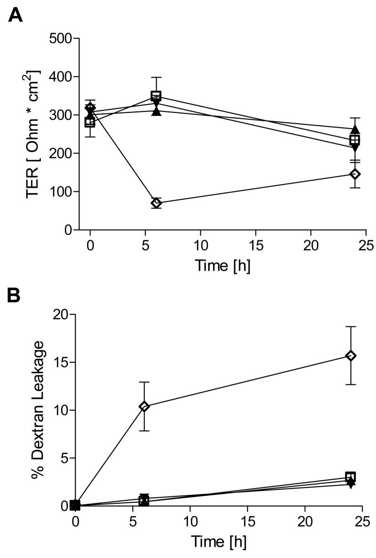
Figure 2. Exposure to DETA NONOate does not cause barrier dysfunction in 16HBE14o- cells.
Effects of the indicated concentrations of DETA NONOate (100 and 500 μM) on (A) transepithelial electrical resistance and (B) FITC-dextran leakage were monitored in 16HBE14o- cells over 24 hrs. For comparison, loss of TER and increased FITC-dextran permeability upon TJ disruption by the Ca2+-chelator EGTA (2 mM) are shown (n=4). □ = control; ▴= DETA–NO 100 μM; ▾ = DETA – NO 500 μM; ⋄ = EGTA 2mM.

