Figure 3.

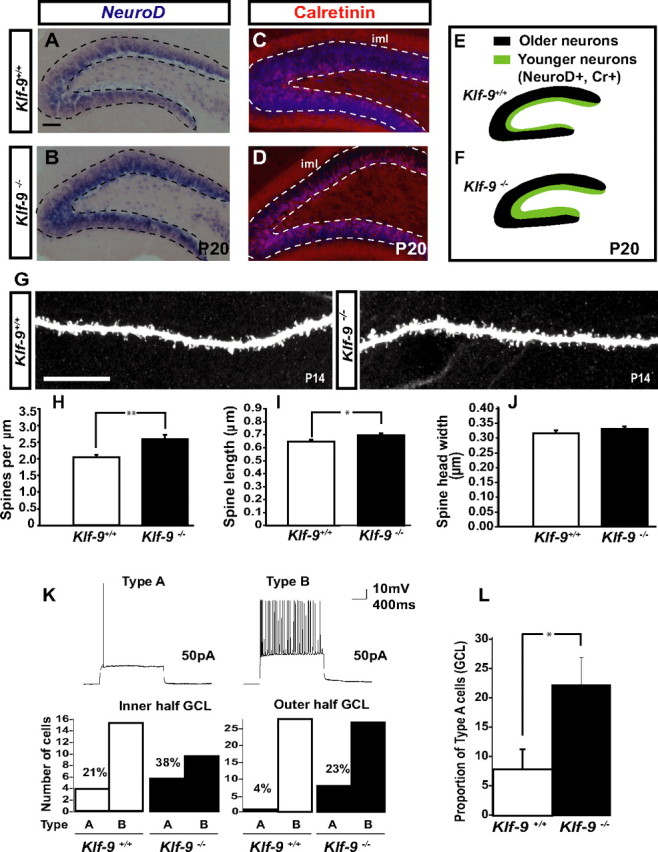
Dentate granule neurons lacking Klf-9 show prolonged expression of early-phase markers, have longer dendritic spines, and exhibit altered action potential response properties during DG development. A, B, In situ hybridization for NeuroD showing an expansion in the NeuroD population in the DG of P20 Klf-9−/− mice (B) compared with Klf-9+/+ mice (A). C, D, Immunohistochemistry for calretinin showing an expansion in the population of calretinin-expressing cells in P20 Klf-9−/− mice (D) compared with Klf-9+/+ mice (C). E, F, Schematic summarizing NeuroD and calretinin expression in the DG of Klf-9+/+ mice (E) and Klf-9−/− mice (F) at P20; n = 3 mice/genotype. G, Representative image of a luciferase-filled dendrite of a Klf-9+/+ (left) and Klf-9−/− (right) dentate granule neuron at P14. H–J, Quantification of spine density, Klf-9+/+, 2.08 ± 0.08 spines per micrometer; Klf-9−/−, 2.64 ± 0.13 spines per micrometer; p = 0.002, unpaired t test. I, Spine length, Klf-9+/+, 0.66 ± 0.012 μm; Klf-9−/−, 0.71 ± 0.01 μm; p = 0.019, unpaired t test. J, Spine head width, Klf-9+/+, 0.32 ± 0.01 μm; Klf-9−/−, 0.34 ± 0.008 μm; p > 0.05, unpaired t test; n = 3 mice per genotype. K, L, Based on AP response properties of dentate granule neurons (P14–P17) in response to current injection (3 s; 50 pA), cells in DG were classified as “type A” or “type B.” Of cells in the inner one-half of the DG, 38% are type A in Klf-9-null mice compared with 21% in controls. Twenty-three percent of cells in the outer one-half of DG are type A in Klf-9−/− mice in contrast to only 4% in controls. The DG of Klf-9−/− mice have a significantly greater proportion of type A cells than controls (L) (p = 0.029, unpaired t test; n = 9 mice per group). Results are mean ± SEM. Scale bar: A–D, 100 μm; G, 10 μm. *p < 0.019 (I); **p < 0.002 (H).
