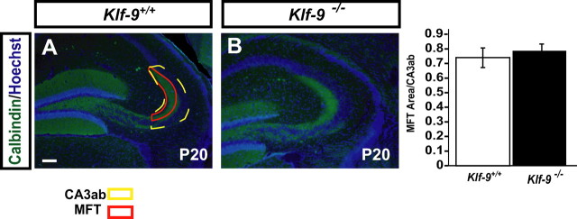
Figure 4.
Mossy fiber projections develop normally in Klf-9-null mice. A, Representative micrograph of calbindin immunohistochemistry showing normal mossy fiber targeting in Klf-9−/− mice (right) compared with Klf-9+/+ mice (left) at P20. B, Mossy fiber terminal area is not different between Klf-9+/+ and Klf-9−/− mice (p > 0.05, unpaired t test); n = 3 mice per group. Results are mean ± SEM. Scale bar, 100 μm.

