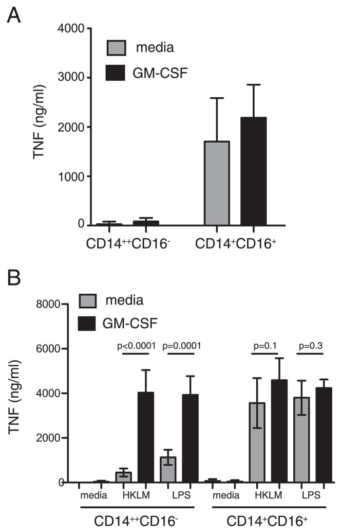
FIGURE 4.
Effect of GM-CSF priming on TNF secretion by monocytes following microbial stimulation. CD14+CD16− and CD14+CD16+ monocytes were purified from flow through fractions of 4–5 donors and cultured with GM-CSF or medium alone for 16 h. At the end of culture, GM-CSF was removed, fresh medium was added, and cells were infected with underminated conidia (A) or stimulated with HKLM or LPS (B). Supernatants were removed 16 h later, and TNF-α secretion was analyzed by ELISA. Shown are mean values (error bars, SD). The unpaired Student t test was used to compare groups. Statistical analysis was performed with Prizm 5. A value of p = 0.05 was considered significant.

