Figure 3.
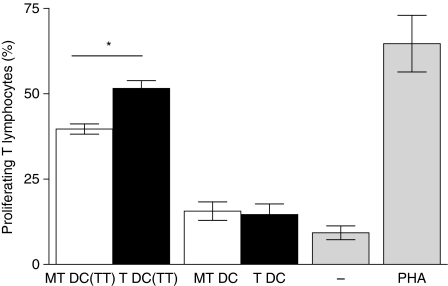
Increased T-lymphocyte proliferation in mixed leucocyte reactions with neuraminidase-treated dendritic cells (DCs). Human monocyte-derived DCs (moDCs) were stimulated with tetanus toxoid (TT; 5 μg/ml), inactivated with mitomycin C and treated [T DC(TT)] or mock-treated (i.e. inactivated neuraminidase) [MT DC(TT)] with neuraminidase, as described in the Materials and methods. Control assays with unstimulated moDCs, treated (T DC) or mock-treated (MT DC) with neuraminidase, were also performed in parallel. The moDCs were then cultured with autologous, carboxyfluorescein diacetate succinimidyl ester (CFSE)-labelled CD14− peripheral blood mononuclear cells (PBMCs; 1 : 8 ratio), for 7 days. As a control, CD14− PBMCs were cultured alone, in the absence or presence of phytohaemagglutinin. Cells were stained with allophycocyanin-labelled anti-CD3 and analysed by flow cytometry. Graph represents the percentage of proliferating T lymphocytes, of five individual experiments ± SEM, estimated by the percentage of CFSE-diluted, CD3+ cells. Significantly different values were observed for the percentage of T-lymphocyte proliferation induced by T DC(TT) (*P< 0·05 paired Student’s t-test), when compared with the one induced by MT DC(TT).

