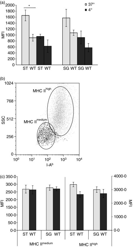
Figure 5.
Major histocompatibility complex (MHC) expression is increased in ST3Gal.I−/− and ST6Gal.I−/− bone marrow-derived dendritic cells (BMDCs). The MHC II expression of ST3Gal.I−/− (ST) and ST6Gal.I−/− (SG) BMDCs was analysed by staining with phycoerythrin-conjugated anti I-Ab, after exposure to fluorescein isothiocyanate-conjugated ovalbumin and incubation at 37° or 4°, as described in the Materials and methods. Control assays were performed in parallel with wild-type (WT) BMDCs. (a) The results are expressed as the mean fluorescence intensity (MFI) ± SEM of ST3Gal.I−/− BMDCs (six mice) and ST6Gal.I−/− BMDCs (seven mice). Significantly different values, related to WT BMDC, were observed at 37°, for the ST3Gal.I−/− BMDCs (*P< 0·05). (b) Dot-plot showing that, after endocytosis (at 37°), BMDCs express different levels of MHC II, defining a MHC IImedium and a MHC IIhigh subpopulation. The data shown correspond to analysis of one representative population of BMDCs (either from WT, ST3Gal.I−/− or ST6Gal.I−/− mice) acquired by flow cytometry. (c) MHC IImedium subpopulations of ST3Gal.I−/− and ST6Gal.I−/− BMDCs are no different from WT BMDCs, while MHC IIhigh subpopulations showed increased MHC II expression. The results are the means ± SEM of the MFI for MHC II for the MHC IImedium and MHC IIhigh subpopulations, gated as described above. The left axis corresponds to the MHC IImedium subpopulation and the right axis corresponds to the MHC IIhigh population.

