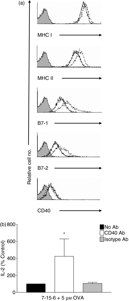
Figure 8.
Incubation of immunogenic L1210 clones with anti-CD40 antibodies (Abs) increases antigen-presenting cell (APC) function. (a) Immunogenic L1210 clone 7-15·6 was incubated for 24 hr with 10 μg/ml anti-CD40 Ab and the surface expression of major histocompatibility complex class I (MHCI), MHCII, B7-1, B7-2 and CD40 was examined by flow cytometry. Shaded histograms represent isotype control staining, solid lines indicate surface expression in untreated 7-15·6 cells, and dotted lines represent surface expression after anti-CD40 Ab treatment. Histograms are representative of two independent experiments performed. (b) Clone 7-15·6 was pulsed overnight with whole ovalbumin (OVA; 5 μm) and incubated either in the absence of Ab (black bars), with 10 μg/ml anti-CD40 Ab (white bars), or with isotype-matched control Ab (grey bars), and combined with primary DO11·10 T cells at an effector-to-target (E:T) ratio of 1 : 1. Supernatants were collected and subsequently assayed for interleukin (IL)-2 by enzyme-linked immunosorbent assay (ELISA). IL-2 stimulated by cells in the absence of Ab is represented as 100%. Data represent the percentage of IL-2 (mean± standard deviation) stimulated in three independent experiments performed; *, P< 0·05, compared with isotype-treated controls.

