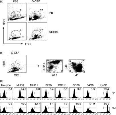
Figure 1.
Phenotypic characteristics of granulocyte colony-stimulating factor (G-CSF)-induced immature myeloid cells (gMCs). Naïve B6 mice were injected subcutaneously with control phosphate-buffered saline (PBS) or G-CSF (10 μg) daily for 5 days. Peripheral blood cells (PBCs) and plenocytes were obtained and the subpopulation of cells was analysed by flow cytometry. (a) The dot plot analysis shows the percentage of gated cells analysed for forward-scatter (FSC) and side-scatter (SSC) properties. (b) Splenocytes were obtained from G-CSF-injected B6 mice and stained with fluorscein isothiocyanate (FITC)-anti-Gr-1 and phycoerythrin (PE)-anti-CD11b or FITC-anti-linage markers and PE-anti-c-kit. Data represent the percentage of quadrant gated on R1. (c) Splenocytes (SP) and bone marrow cells (BM) were obtained from G-CSF-injected and naïve B6 mice, respectively. Cells were stained for Gr-1, CD11b, and each surface marker was investigated by three-colour fluorescence. The histogram shows the percentage of positive cells gated from the Gr-1+ CD11b+ cells. Representative data from one of three experiments are shown; all three experiments showed similar results.

