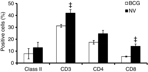
Figure 4.
Phenotypic analysis of guinea pig lung digest cells. Lung cells obtained by enzymatic digestion from non-vaccinated (NV) and bacillus Calmette–Guérin-vaccinated (BCG) guinea pigs 5 weeks after infection with virulent Mycobacterium tuberculosis was characterized by flow cytometry. Proportions of major histocompatibility complex (MHC) class II+ cells, T cells and their subsets were determined by fluorescence-activated cell sorting analysis after staining the cells with monoclonal antibodies directed against the surface markers of MHC class II+ cells, T cells (CT5), CD4+ T cells (CT7), and CD8+ T cells (CT6). Results are expressed as % positive cells. ‡P< 0·01 compared with BCG-vaccinated group as determined by Student’s t-test.

