
Figure 3.
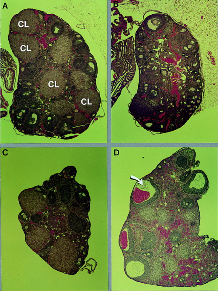
Morphology of normal and C/EBPβ-deficient ovaries from untreated animals or after treatment with gonadotropins in vivo. Photomicrographs (40×) of sections of hematoxylin/eosin-stained ovaries from heterozygous (A) and homozygous (B) mutant ovaries of 2- to 3-month old mice, and of heterozygous (C) and mutant (D) ovaries from mice treated for 2 days with PMSG and subsequently for 14 hr with hCG (see Table 2). The arrow indicates an oocyte that has failed to be expelled. (CL) Corpus luteum.