
Figure 4.
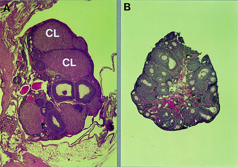
Morphology of transplanted ovaries. Photomicrographs (50×) of sections of hematoxylin/eosin-stained ovaries from a heterozygous ovary (A) 3 months after transplantation into a homozygous mutant host (see Table 3A) and a mutant ovary (B) 2 months after transplantation into a wild-type host animal (see Table 3B). (CL) Corpus luteum.