
Figure 5.
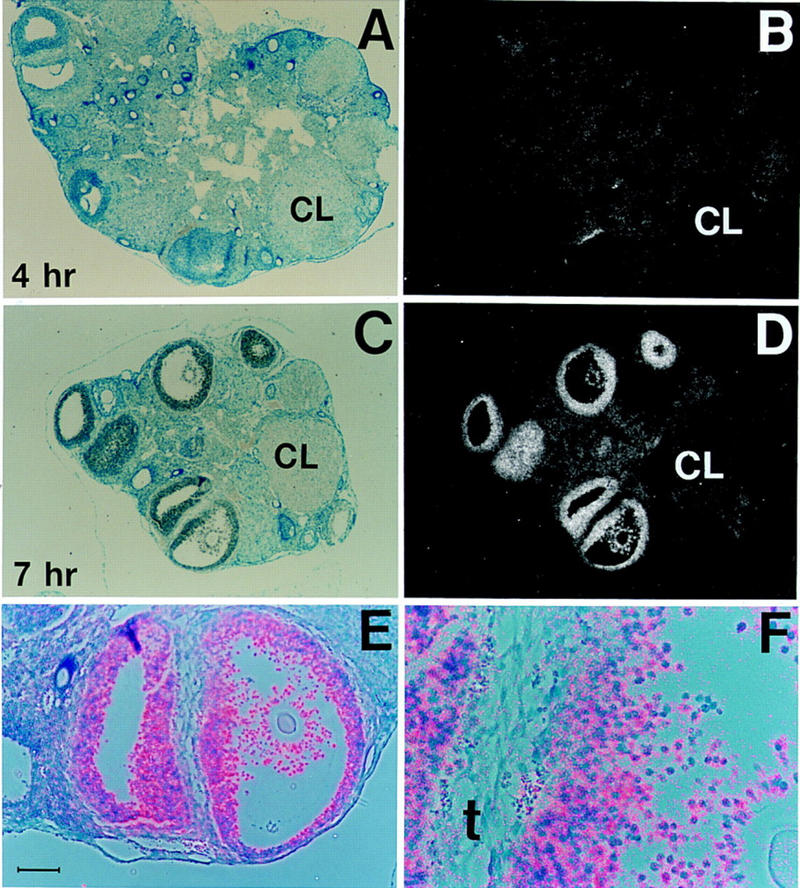
C/EBPβ expression is induced in granulosa cells 4–7 hr after hCG treatment. Bright-field (A,C), dark-field (B,D), and bright- plus dark-field (E,F) photomicrographs of sections from normal adult ovaries after in situ hybridization to a C/EBPβ-specific antisense cRNA probe. The animals were treated for 2 days with PMSG and then with hCG for 4 hr (A,B) and 7 hr (C,D), respectively. E and F show high magnification of the two follicles shown at the bottom of C and D; silver grains (predominantly over granulosa cells) are in pink. The scale bar in E represents 10 μm. (A,B) C/EBPβ+/− ovaries; (C–F) C/EBPβ+/+ ovaries. (CL) Corpus luteum; (t) theca layer. Magnifications, 62.5× (A–D); 100× (E); 320× (F).