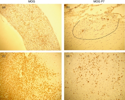
Figure 2.
Pathology of optic nerve (a, b) and spinal cord (c, d) lesions in rats that had been immunized with myelin oligodendrocyte glycoprotein (MOG) (a, c) and MOG peptides (P7 and P8) (b, d). Histological examinations revealed relatively mild pathology in MOG peptide-immunized rats. ED1 staining for macrophages demonstrated extensive macrophage infiltration in the optic nerve (a) and spinal cord (c) of MOG-immunized rats and mild and localized (dotted line) macrophage infiltration in MOG peptide-immunized rats (b, d). (a, b) MOG immunization, day 14; (c, d) MOG peptides (P7 + P8) immunization, day 12. (a) × 60; (b–d) × 120.

