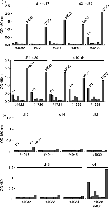
Figure 4.
Kinetics of anti-myelin oligodendrocyte glycoprotein (anti-MOG) (a) and anti-MOG peptide (b) antibodies in MOG-immunized rats. The levels of anti-MOG and anti-MOG peptide antibodies were measured in triplicate by enzyme-linked immunosorbent assay. Recombinant MOG or MOG peptides (P1–P8) (10 μg/ml) were coated onto microtitre plates and diluted sera (1 : 100) from MOG-immunized rats were applied. After washing, horseradish peroxidase-conjugated anti-rat immunoglobulin G was allowed to react. The reaction products were then visualized after incubation with the substrate and the absorbance was read at 450 nm. During the early stage (day 14–21), anti-MOG antibodies were elevated, while anti-MOG peptide antibodies were not detected. From day 32 antibodies against P1 were also detected. All SDs were within 10% of the mean values.

