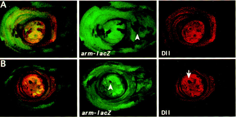
Figure 2.

Mitotic recombinant clones in leg discs induced by the FRT–FLP system at 72–96 hr AEL (early third instar). Leg discs were stained with anti-β-gal (green) and anti-Dll (red). (A) Wild-type clones marked by the absence of β-gal expression. The arrowhead indicates a clone that extends to the boundary between Dll expressing and Dll nonexpressing cells. (B) DllSA1 clones marked by the absence of β-gal and Dll expression and their twin spots by the elevated level of β-gal. Note that the DllSA1 clones do not proliferate further. Here and in all remaining images of leg discs, dorsal is to the right.
