
Figure 3.
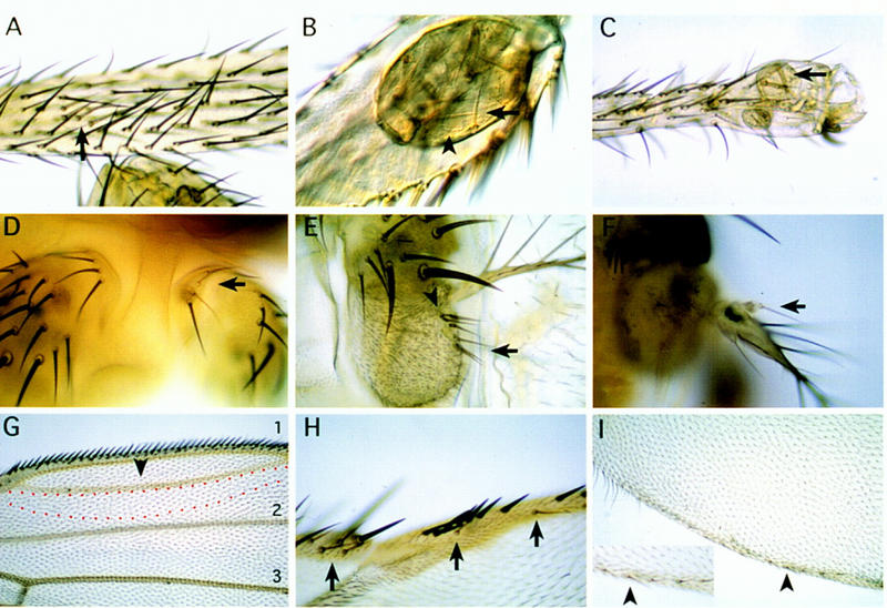
Phenotypic effects of DllSA1 clones in the leg (A–C), antenna (D–F), and wing (G–I). The clones were induced during 24–120 hr AEL and marked with y except for G where clones were marked with forked36(f36) (see Materials and Methods). (A–C) Clones in the leg. Early induced clones (24–48 hr AEL) only appear in the coxa as it has been described previously. Clones induced later (72–120 hr AEL), however, are able to proliferate and differentiate nonbracted bristles in the proximal tibia (A) and vesicles of y− tissue that segregate from the surrounding wild-type tissue in the distal tibia (B) and tarsus (C). Arrows indicate y bristles; arrowheads indicate trichomes that are not present in the distal leg. (D–F) Clones in the antenna. An early (24–48 hr AEL) clone in aI (D) does not produce a mutant phenotype as aI does not require Dll activity. Late clones (72–120 hr AEL) in the aIII antennal segment (E) and arista (F) develop bristles sometimes with an associated bract. Arrows indicate y− bristles; arrowheads indicate bracted bristles. (G–I) Clones in the wing. The clones near the D/V margin give rise to extra-vein tissue. The red dashed line indicates a dorsal clone marked with f close to vein 1. Normal veins 1, 2, and 3 are indicated. Arrowhead indicates extra-vein (G). Clones that abut the D/V boundary also eliminate bristles of the triple row in the A compartment (H) and long hairs of the double row in the P compartment (I). (Inset in I) Magnification showing the y− bristles with socket in the P compartment (arrowhead). Arrows indicate y bristles.