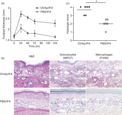
Figure 2.
Enhanced leucocyte recruitment after antigen-dependent conditioning in a peptide delayed type hypersensitivity (DTH) model. (a) The DTH reaction was induced by transfer of antigen-specific pioneer Th1 cells into recipient mice followed by s.c. injection of ovalbumin (OVA)323–339 (OVAp)/incomplete Freund’s adjuvant (IFA) or phosphate-buffered saline (PBS)/IFA into the footpad. Footpad swelling was determined over time (n = 5). (b) Representative histological staining [haematoxylin and eosin (H&E); myeloperoxidase (MPO) for granulocytes and F4/80 for macrophages; magnification ×200] of OVAp/IFA- and PBS/IFA-injected footpads 24 hr after antigen application. (c) A summary of individual histological evaluations according to the histological score of infiltration and tissue destruction of OVAp/IFA- and PBS/IFA-injected sites (score grade 0, normal tissue; score grade 1, loose infiltrates in subcutaneous tissue; score grade 2, moderate, predominately phlegmonous infiltrates in subcutaneous tissue; score grade 3, abscess formation in subcutaneous tissue; score grade 4, abscess formation with infiltration of skeletal muscle). *P < 0·05.

