Figure 3.
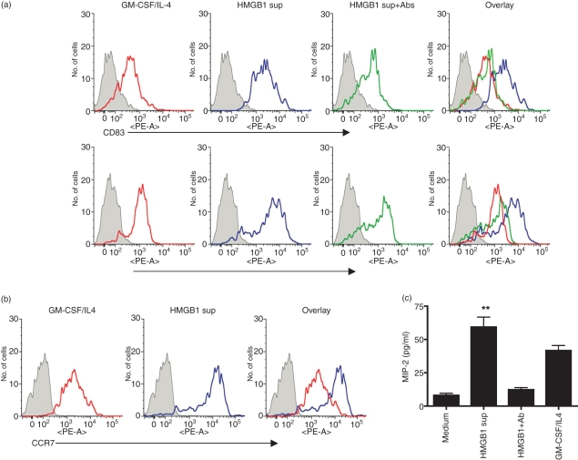
High-mobility group box 1 (HMGB1) induces the phenotypic maturation of dendritic cells (DCs). Fluorescence-activated cell sorter (FACS) analysis of immature DC cultured in the presence of granulocyte–macrophage colony-stimulating factor (GM-CSF)/interleukin-4 (IL-4) medium or HMGB1 plasmid-transfected cell supernatant. (a) Bone marrow-derived dendritic cells (BMDC) of BALB/c mice were resuspended in culture medium supplemented with GM-CSF/IL-4 (red line) or in the presence of HMGB1mut plasmid-transfected cell supernatant (HMGB1 sup) (blue line) or cell supernatant depleted with rabbit polyclonal antibody to HMGB1 (HMGB1 sup + Ab) (green line) and cultured for 7 days. On day 7, the cells were harvested, washed and analyzed for surface expression, by flow cytometry, using the indicated markers. (b) Treatment of HMGB1 in up-regulation of chemokine receptor expression. Cells were stained for CCR7 and analyzed using FACS. Histograms show the staining of specific surface markers, and filled histograms represent the isotype-matched control antibody staining. The FACS data are representative of three separate experiments, with similar results obtained on each occasion. (c) Chemokine secretions in response to HMGB1 treatment. The concentration of macrophage inflammatory protein-2 (MIP-2) was determined in the supernatant using enzyme-linked immunosorbent assay (ELISA) analysis. All error bars represent standard deviation (SD) and are representative of three independent experiments. **Significant differences by comparison with the medium control (P < 0.001; Student’s unpaired t-test). PE, phycoerythrin.

