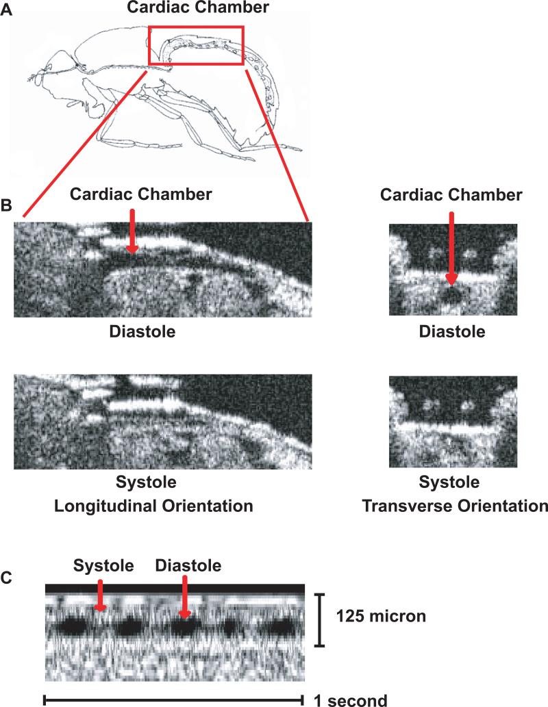
Figure 1.
OCT images of cardiac chamber in adult w1118 Drosophila. (A) Schematic representation of the cardiac chamber (red box) of an adult Drosophila based on Miller [45]. (B) Longitudinal and transverse B-mode OCT images of the cardiac chamber in adult Drosophila during diastole and systole. (C) Representative m-mode OCT image demonstrating the cardiac cycle with diastole and systole denoted by the red arrows.

