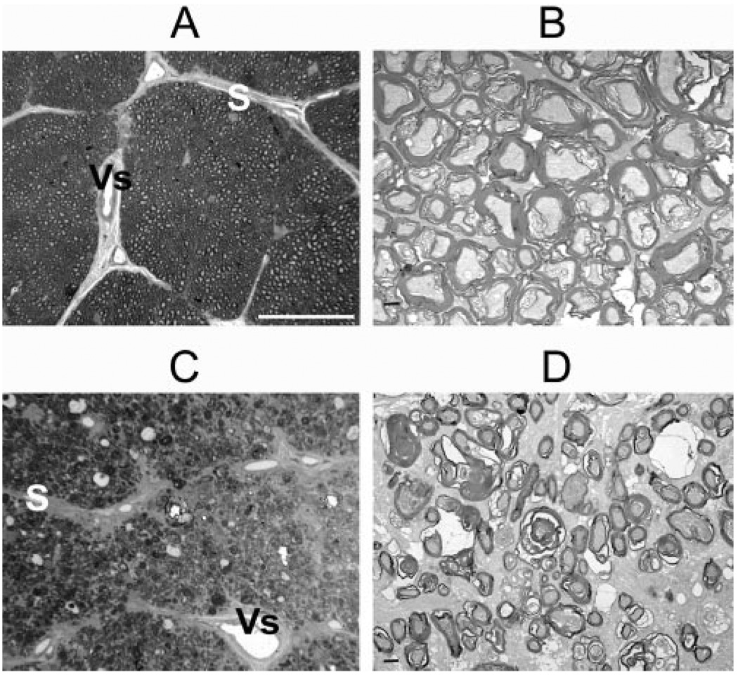
FIGURE 6.
Cross sections of ONs from normal (A, B) and pNAION (C, D) eyes. (A) Toluidine blue–stained section from a control ON showed normal-appearing axon bundles separated by fibrovascular pial septae (S), some of which contained capillaries (Vs). Axons were seen as small, unstained circles surrounded by stained myelin sheaths. (B) TEM section from same area as in (A) showed regular, myelinated axon bundles. (C) Toluidine blue–stained section from a pNAION ON showed loss of axons, reduction in size of axon bundles, and thickening and disruption of fibrovascular pial septae (S). V, central retinal vein. (D) TEM section from same area as (C) showed extensive demyelination with shrinkage and disruption of axons. Scale bars: (A, C) 40 µm; (B, D) 1 µm.

