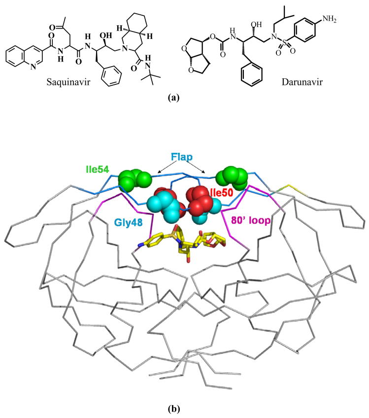
Figure 1.
(a) The chemical structures of saquinavir and darunavir. (b) Structure of HIV-1 PR dimer with the locations of mutated residues Gly48 (cyan), Ile50 (red), Ile54 (green) indicated by spheres for main chain atoms in both subunits. Darunavir is shown in sticks colored by atom type. The flap residues (45–55) and the 80’s loop (78–82) are colored in blue and purple, respectively.

