
Figure 3.
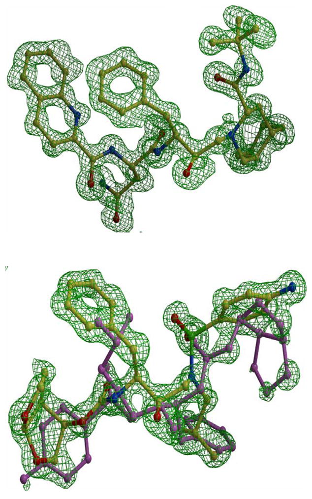
The Fo-Fc omit maps of saquinavir (upper panel) and darunavir (lower panel) contoured at 3.3 sigma. Saquinavir is colored by atom type from complex PRI54V-SQV. Darunavir is from complex PRI54V-DRV showing alternate conformations of 60/40% occupancy. The major conformation is colored by atom type and the minor is colored pink.