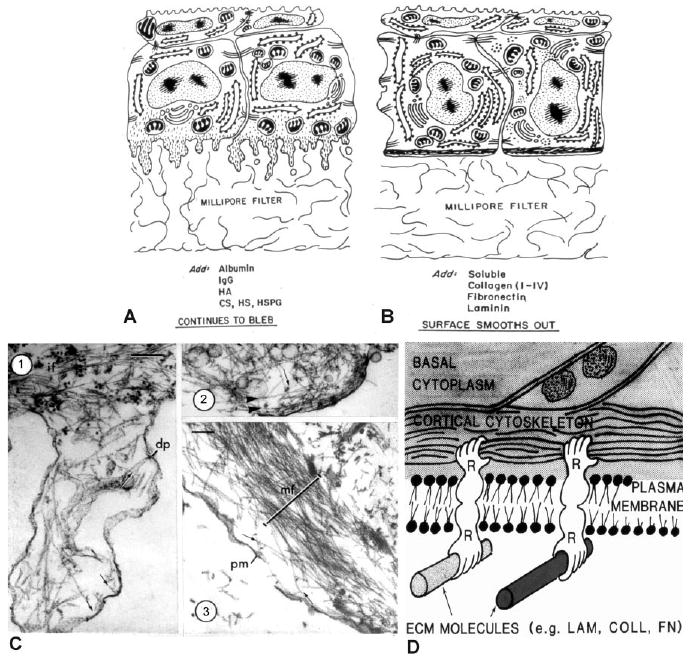
Fig. 2.

Diagrams and electron micrographs illustrating the effects of different experimental conditions on the organization of the basal cell surface. A: The basal surface blebbed when the basal lamina was removed by EDTA or enzyme treatment, and the blebbing persisted on Millipore filters in the presence of nonmatrix proteins or glycosaminoglycans. B: Soluble collagens, fibronectin and laminin added to the medium stimulated the bleb retraction and reformation of the basal actin cytoskeleton. Reprinted from Sugure and Hay (1982). C: Corneal epithelial tissues were detergent extracted and treated with S-1 fragments of heavy meromyosin before EM preparation. C-1 is a typical bleb demonstrating a core of actin filaments decorated with myosin S-1 fragments, which aligns on the actin filaments indicating the polarity of the filament that is pointing toward the plasma membrane (indicated by small arrows) and some apparently inserting into a dense plaque (dp). Intermediate filaments (IF) were in the basal cell area. C-2 is a smaller bleb with some actin filaments parallel to the cell surface (arrowheads) and others perpendicular to the surface. C-3 presents a cell 6 hr after immersion in soluble type IV collagen (100 μg/ml). The actin bundle (MF) was in the basal compartment of this slightly tangential section containing a damaged plasma membrane (PM). The actin filaments were organized in opposite directions (small arrows). Scale bars = 0.2μm. Reprinted from Sugrue and Hay (1982). D. Hay's concept drawing illustrating the influence of the ECM on the actin cytoskeleton. It was used in lab meetings and titled “Hands across the membrane.”
