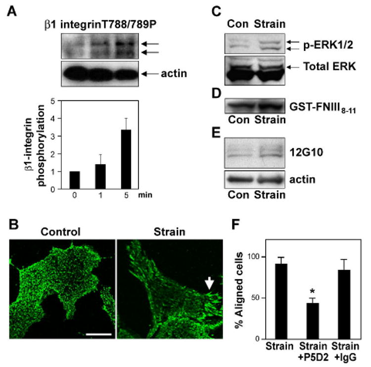
Fig. 2. ·1 integrin activation is required for cyclic strain-induced reorientation of CE cells.

A) Western blot analysis of CE cell lysates showing time-dependent phosphorylation of ·1 integrin cytoplasmic tail at threonine T788/789 in response to static stretch. Histogram shows the corresponding densitometric quantification of ·1 integrin phosphorylation. B) Immunofluorescence micrographs of control and strain-exposed CE cells stained for activated β1 integrin using 12G10 antibody. Arrow indicates increased clustering of activated ·1 integrins within large streak-like focal adhesions at the cell periphery. Scale bar: 25 ·m. c-e) Western blots showing MAP kinase (ERK1/2) phosphorylation (C) and binding of GST-FNIII8-11 (D) and 12G10 (E) in CE cells in the absence and presence of static (C) or cyclic strain (D, E). F) Percentage of cells oriented 90 ± 30 ° degrees (aligned) relative to the direction of applied cyclic strain in the absence or presence of the ·1 integrin blocking antibody P5D2 (p < 0.001) or isotype-matched IgG.
