
Fig. 3. Mechanical strain-induced ·1 integrin activation requires the PI3K/AKT pathway.
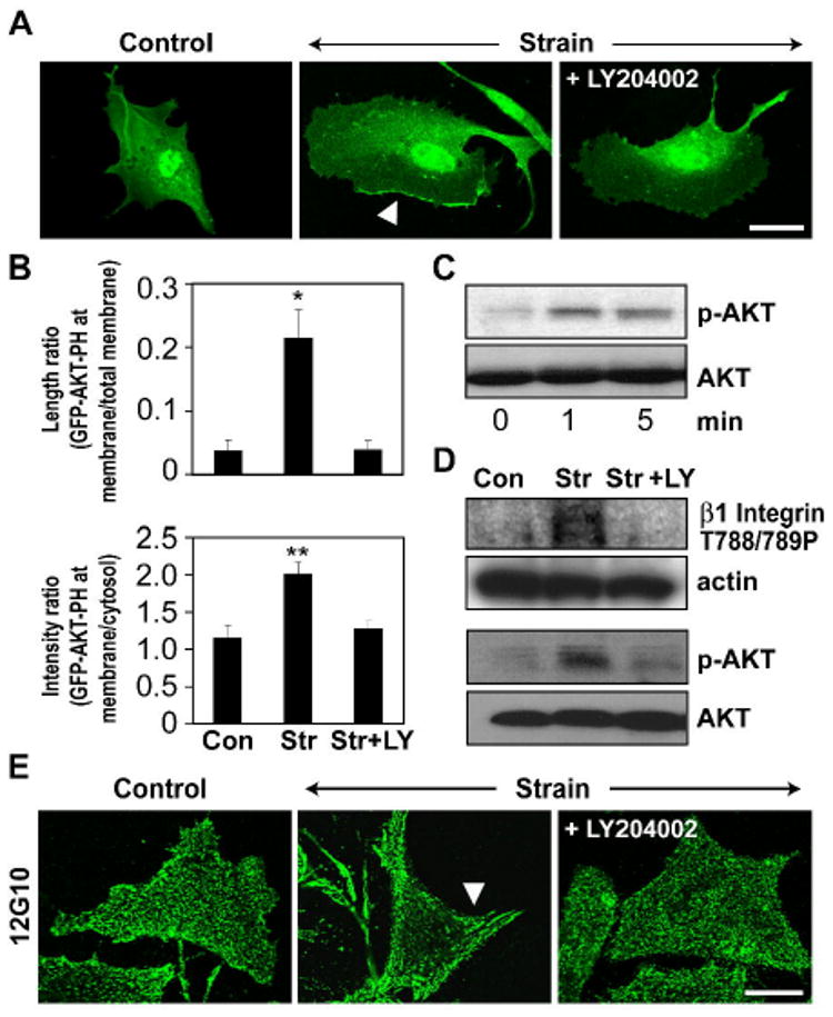
A) Fluorescence micrographs of CE cells transfected with GFP-AKT-PH and subjected to 0 or 15% static stretch in the absence or presence of the PI3-Kinase inhibitor, LY 294002 (LY, 40 ·M). Note that LY 294002 inhibits strain induced translocation of GFP-AKT-PH domain to the plasma membrane (arrow). Scale bar: 25 ·m. B) Quantification of mechanical strain-induced GFP-AKT-PH domain translocation to the membrane in the absence or presence of the PI3K inhibitor LY294002, measured as a fraction of total cell membrane perimeter that is enhanced with GFP-AKT-PH in randomly selected cells and the ratio of GFP fluorescence intensity in the membrane versus cytosol (*, p < 0.05). C-D) Representative Western blots showing time dependent activation of AKT (C) and phosphorylation of ·1 integrin cytoplasmic tail at T788/789 and AKT at ser-473 in response to static stretch in the presence and absence of the PI3K inhibitor, LY294002 (D). E) Fluorescence micrographs of CE cells subjected to 0 or 15% mechanical strain in the absence or presence of the PI3-Kinase inhibitor, LY 294002 and stained for activated ·1 integrin using the12G10 antibody. Arrow indicates increased clustering of activated ·1 integrins within large streak-like focal adhesions at the cell periphery. Scale bar: 25 ·m.