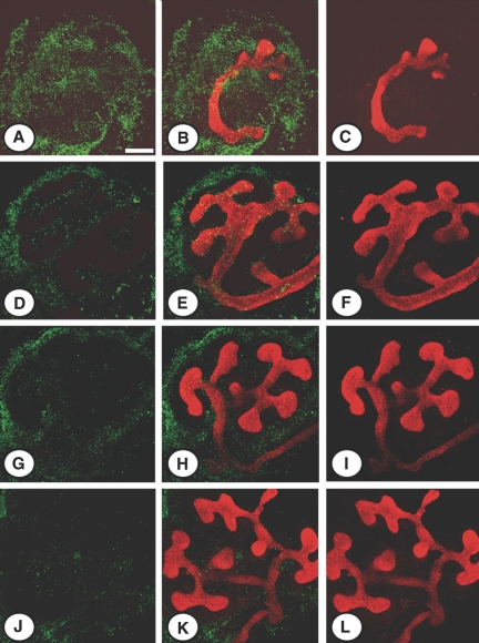
Figure 8.
Effect of sSCF on apoptosis in vitro. Representative photographs of E11.5 kidneys from MMP9−/− (A through F) and MMP9+/+ (G through L) kidneys stained with calbindin D-28K to delineate ureteric bud in red and the TUNEL method (apoptotic nuclei in green). Kidneys were grown for 48 h in the absence (A through C and G through I) or the presence (D through F and J through L) of physiologic concentration of sSCF in culture medium. sSCF in culture medium decreased the number of apoptotic cells and increased ureteric bud branching of MMP9+/+ (J through L versus G through I) and MMP9−/− (D through F versus A through C) kidneys. Bar: 250 μm.

