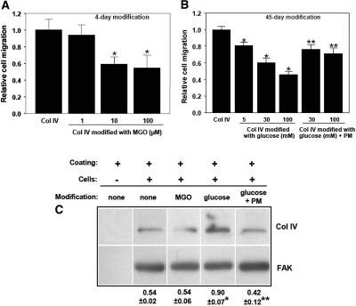
Figure 4.
Cell migration on MGO- or glucose-modified collagen IV and effect of collagen modifications on endogenous collagen IV expression in mesangial cells. Transwells were coated with collagen IV and modified with (A) MGO, and (B) d-glucose or d-glucose and 20-mM PM at 37°C. After washing and blocking, mesangial cells (1 × 10⁴) were plated on the top of Transwell membranes and cell migration was determined after 6 h at 37°C as described in the Concise Methods section. (C) Mesangial cells were plated on unmodified or modified collagen IV for 72 h. The levels of intracellular collagen IV were determined in total cell lysates by Western blot using anti-collagen IV antibody. Membranes were subsequently incubated with anti-FAK antibody to verify equal loading. Collagen IV and FAK bands were quantified by densitometry analysis, and the collagen IV signal was expressed as the collagen IV-to-FAK ratio. *P < 0.05 (n = 3), MGO versus no MGO, or glucose versus no glucose; **P < 0.05 (n = 3), glucose + PM versus glucose.

