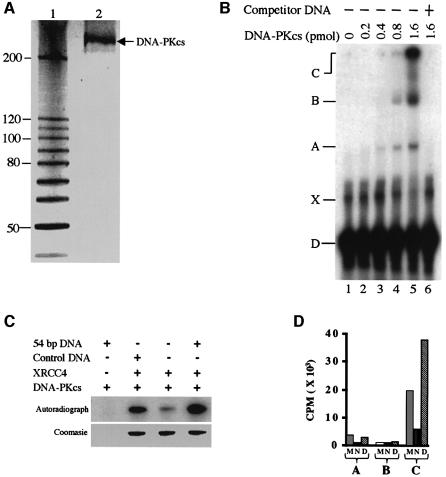
Fig. 1. Purification and functional assays of DNA-PKcs. (A) Silver-stained SDS–gel of purified human DNA-PKcs. Lane 1, 10 kDa ladder protein marker (Gibco); lane 2, purified DNA-PKcs. (B) EMSA of DNA-PKcs bound to 54 bp DNA. 5′-32P-labelled dsDNA (20 fmol) was titrated with an increasing amount of DNA-PKcs. In lane 6, 500 ng of unlabelled 54 bp DNA were added as competitor. (C) Autoradiography of DNA-dependent kinase activity over XRCC4. Protein bands correspond to the XRCC4 in the experiment. Control DNA, 100 bp ladder (New England BioLabs). (D) Radioactivity measurement of DNA-dependent kinase activity on DNA-PKcs (autophosphorylation) (lanes A and B) and XRCC4 (lane C). Protein bands obtained in experiments similar to those shown in ‘C’ were cut and the radioactivity was measured by Cerenkov counting. CPM, counts per minute; A, autophosphorylation of DNA-PKcs in the absence of XRCC4; B, autophosphorylation of DNA-PKcs in the presence of XRCC4; C, phosphorylation of XRCC4; M, phosphorylation in the presence of control DNA; N, phosphorylation in the absence of DNA; D, phosphorylation in the presence of 54 bp DNA.

An official website of the United States government
Here's how you know
Official websites use .gov
A
.gov website belongs to an official
government organization in the United States.
Secure .gov websites use HTTPS
A lock (
) or https:// means you've safely
connected to the .gov website. Share sensitive
information only on official, secure websites.
