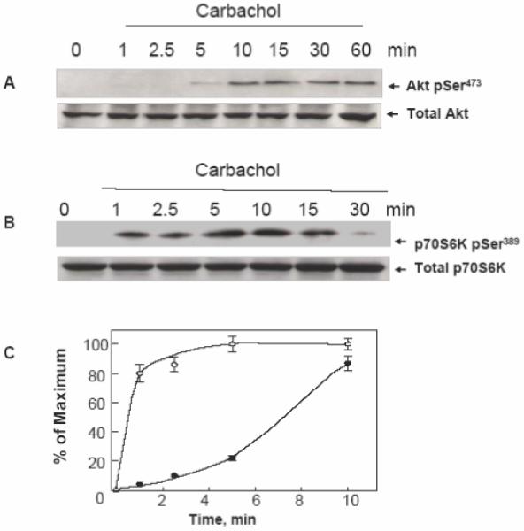
Figure 1. Carbachol induces Akt phosphorylation on Ser473 and p70S6K phosphorylation on Ser389in a time-dependent fashion in T84 cells.

Cultures of T84 cells grown on 35 mm dishes were stimulated with 100 μM carbachol for the indicated times. All cultures were lysed with 2×SDS-PAGE sample buffer and analyzed by SDS-PAGE and immunoblotting with antibodies that detect either Akt phosphorylated on Ser473 (panel A) or p70S6K phosphorylated on Ser389 (panel B). Antibodies that detect total Akt or p70S6K were used to verify equivalent loading of the gel. Autoluminograms were quantified by densitometric scanning. The results shown are the mean ± S.E.M. n=3 and are expressed as percentage of the maximum increase induced by treatment with carbachol for either AKT pSer473 closed symbols or p70S6K pSer389 open symbols (panel C).
