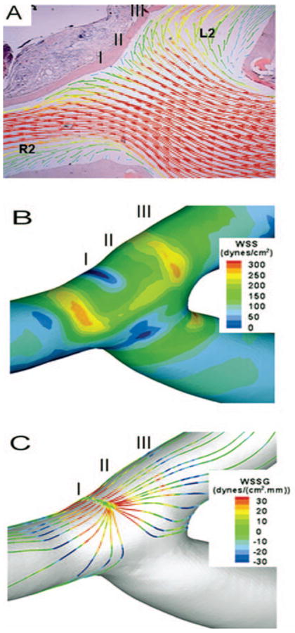
FIGURE 3.

Mapping of hemodynamics. A, calculated velocity field on the plane overlaid on a histological image; vectors indicate the velocity direction and magnitude; colors also indicate velocity magnitude. B, surface distribution of luminal WSS. C, surface distribution of luminal WSSG along the flow streamlines. R2, distal segment of the right CCA; L2, distal segment of the left CCA.
