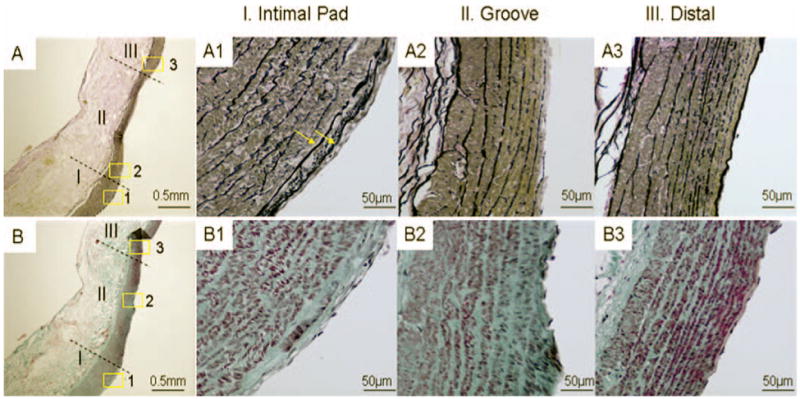
FIGURE 4.

Histological images of impingement (I), acceleration (II), and recovery (III) regions on the left branch of bifurcation 2 months after creation. A–A3, van Gieson staining. B–B3, trichrome staining. A and B, gross morphology showing an intimal pad bordering a “groove” (original magnification, ×40). Insets 1 to 3 in A and B are shown at a higher magnification (×400) in A1 to A3 and B1 to B3. A1 and B1, more elastin deposition (A1, arrows; multiple layers of internal elastic lamina) and more collagen deposition (B1) within the intima hyperplasia in region I, indicating a more mature intimal pad compared with the bifurcation created at 2 weeks (data not shown). The “groove” in region II is characterized by disrupted endothelium and internal elastic lamina and a thinned media (A2) with smooth muscle cell depletion (B2). These are indications of early aneurysm. A3 and B3, region III (distal region on the left branch) shows normal morphology similar to that observed in the vessel before surgery (not shown).
