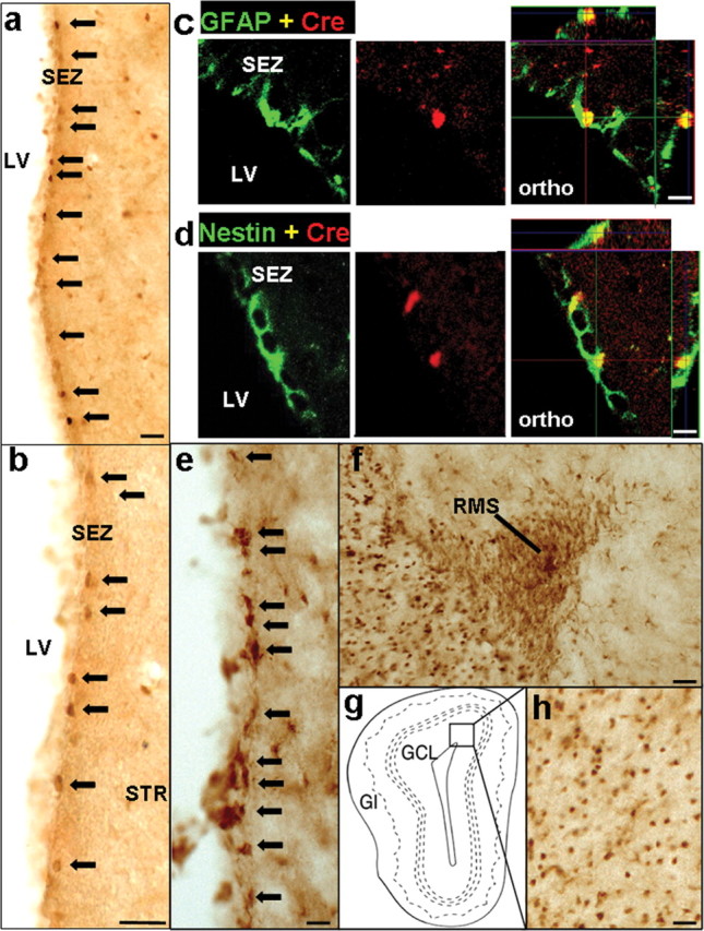
mGFAP-Cre line for conditionally targeting adult neural stem/progenitor cells. a, b, e, f, h, Survey images of coronal sections of the SEZ (a, b, e), RMS (f), and OB (h) stained by bright-field immunohistochemistry. A small number of cells in the SEZ express Cre (a, low mag; b, high mag). Scale bars, 80 μm. c, d, Confocal micrographs of single optical slices through cells in the SEZ that are double stained by immunofluorescence for Cre and GFAP (c) or Cre and Nestin (d). Individual channels and orthogonal analysis show that all Cre-expressing cells in the SEZ also express GFAP (c) and Nestin (d). Orthogonal images (ortho) show three-dimensional analysis of individual cells at specific sites marked by intersecting x, y, and z axes. Scale bars, 20 μm. Many cells express the reporter protein β-gal (e) in the neurogenic proliferative regions of the SEZ. Migrating neuroblasts in the RMS (f) and granule neurons in the OB (h) also express β-gal. Box in g indicates the region shown in h. Arrows in a, b, and e indicate representative Cre or β-gal-positive cells in SEZ. SEZ, Subependmyal zone; LV, left ventricle; STR, striatum; RMS, rostral migratory stream; OB, olfactory bulb; GCL, granule cell layer; Gl, glomeruli.
