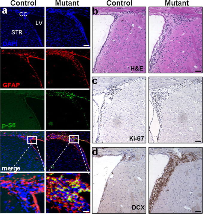
Figure 3.

mGFAP-Cre-mediated Pten deletion leads to expansion of adult neural stem cells and their progenies in vivo. Survey images of coronal sections of control and mutant SEZ. a, Compared with littermate controls, mutant mice showed increased GFAP and P-S6-positive (labeled cells which co-localized in the SEZ. DAPI counter stain is used to visualize nuclei. Scale bars: top, 150 μm; bottom, 25 μm. b–d, Images of H&E (b), Ki-67 (c), and DCX (d) expression demonstrate increased proliferation in mutant SEZ compared with control regions. Scale bar, 60 μm. n = 5. SEZ, Subependymal zone; LV, lateral ventricle; CC, corpus callosum; STR, striatum; H&E, hematoxylin and eosin; DCX, doublecortin.
