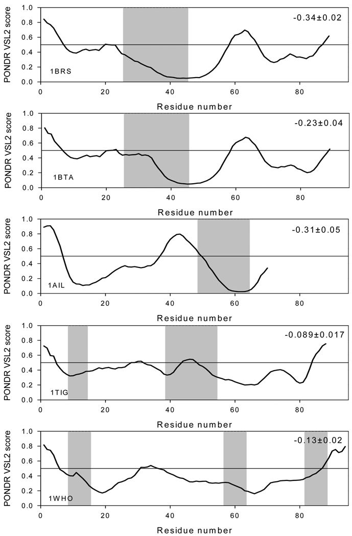
FIGURE 9.
PONDR VSL1 score distributions for proteins in the barnase–barstar-analogue complexes. Localizations of the barstar-binding regions are indicated as gray shaded areas. The values of the mean distance from the 0.5 boundary for each binding region are indicated in the corresponding plot. In PONDR plot, segments with scores above 0.5 correspond to the disordered regions, whereas those below 0.5 correspond to the ordered regions.

