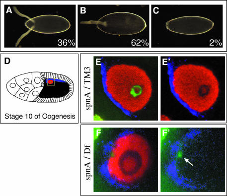
Fig. 1. spnA mutant phenotypes. (A–C) Eggshell phenotype. (A) Wild-type with two anteriorly located dorsal appendages. (B) Mild ventralization with a single fused-dorsal appendage. (C) Completely ventralized. Percentages represent the phenotypic proportions observed per total eggs laid (n = 374) from spnA155–52 germline clones. (D–F′) Gurken expression and karyosome phenotype. (D) Diagram of a wild-type Stage 10 egg chamber. The oocyte nucleus is indicated in red, Gurken protein in blue, karyosome in green. (E and E′) A confocal section of a germinal vesicle from a spnA093A heterozygote Stage 10 egg chamber. Staining for the centrosomal protein, CP190 (red; Whitfield et al., 1995), shows the nucleoplasm. (F and F′) Germinal vesicle from a spnA093A/Df(3R)X3F Stage 10 egg chamber. (E′) is identical to (E) minus the green channel. (F′) is identical to (F) minus the red channel. The yellow box in (D) represents the area captured in (E) and (F). [(A–C) dark-field photographs using 20× objective, anterior to the left; (E and F) confocal images using a 40× objective and 4× zoom, dorsal view with anterior to the left].

An official website of the United States government
Here's how you know
Official websites use .gov
A
.gov website belongs to an official
government organization in the United States.
Secure .gov websites use HTTPS
A lock (
) or https:// means you've safely
connected to the .gov website. Share sensitive
information only on official, secure websites.
