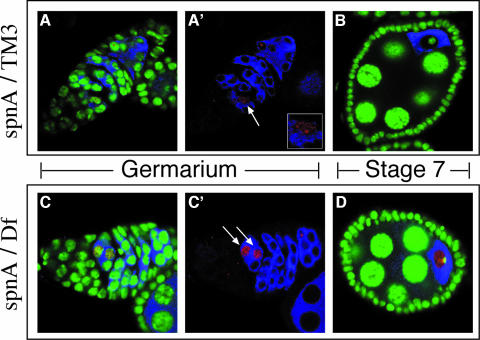
Fig. 7. Persistence of γ-HIS2AV staining suggests a defect in the repair of meiotically induced DSBs. (A and B) Confocal images of different oogenesis stages from a wild-type (spnA093A/TM3) ovary. (C and D) Confocal images of similar oogenesis stages from a spnA [spnA093A/Df(3R)X3F] ovary. DNA is stained with OliGreen® (green). Early cysts and the oocytes are marked by immunostaining for Orb protein (blue). (A and A′) γ-HIS2AV speckles the DNA in one cell (A′, red, arrow), presumably the oocyte. A 2× zoom of that nucleus is provided in the A′ inset. (C and C′) γ-HIS2AV observed in the germarium of spnA ovaries. (C′, arrows) Two cells of a common cyst (D) γ-HIS2AV in spnA mutants beyond the germarium in the vitellogenic stages of oogenesis. All images are single confocal sections.

An official website of the United States government
Here's how you know
Official websites use .gov
A
.gov website belongs to an official
government organization in the United States.
Secure .gov websites use HTTPS
A lock (
) or https:// means you've safely
connected to the .gov website. Share sensitive
information only on official, secure websites.
