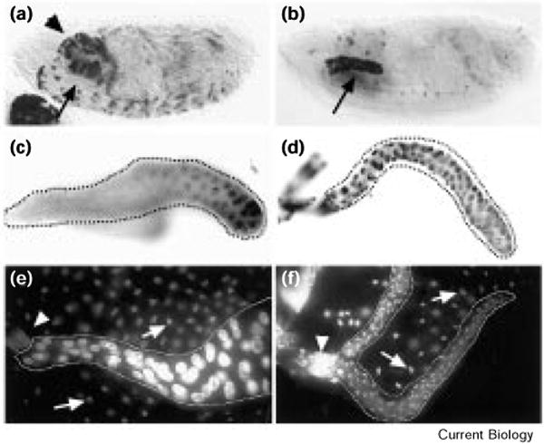
Figure 1.

Continuous Cyclin E expression begins in embryonic salivary glands and continues throughout larval development. (a,b) Embryos and (c,d) salivary glands (outlined) of early third instar larvae were hybridized with a digoxigenin-labeled Cyclin E RNA probe that was visualized using alkaline-phosphatase-conjugated anti-digoxigenin antibodies. Cyclin E RNA accumulated in the salivary glands (arrows) of 43B-UAS–Cyclin E embryos (b) by stage 15, at which time wild-type (Sevelin) embryos (a) do not express Cyclin E in the salivary glands. The arrowhead in (a) indicates labeling in the brain, which is somewhat out of focus in (b). (c) A fraction of the cells of the larval salivary gland express Cyclin E RNA in the wild-type (yw67) salivary glands, whereas all of the cells of the 43B-UAS–Cyclin E glands (d) express Cyclin E. (e) Late third instar wild-type and (f) 43B-UAS–Cyclin E salivary glands (outlined by dotted lines) stained with 1 μg/ml Hoechst 33258. Whereas the salivary glands normally increase in size dramatically during the third instar, the salivary glands of the 43B-UAS–Cyclin E larvae remain small. Arrowheads indicate the ring of diploid cells forming the imaginal ring. Arrows indicate the nuclei of the fat body; cells of the fat body (which are not expressing Cyclin E continuously in these experiments) also endoreplicate but to a lesser extent than cells of the wild-type salivary gland. Note the relative size of the fat body nuclei compared to the wild-type and 43B-UAS–Cyclin E salivary gland nuclei.
