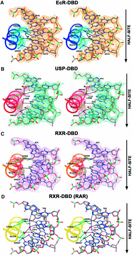
Fig. 3. Stereo views of the recognition α-helices and their contacts to DNA. Shown are interactions with the major grooves for (A) EcR (blue), (B) USP (red), (C) RXR (gold) with a composite omit electron density map (2Fo – Fc) shown for the side chains and the DNA base pairs, and (D) RXR from the RAR–RXR/DR-1 complex (yellow). In all the cases, the side chains that make direct or water-mediated contacts to the bases are shown in green, the water molecules that mediate the contacts between the protein and DNA are yellow/magenta spheres, and hydrogen bonds are shown as dotted lines. The orientation of the half-site is indicated by arrows.

An official website of the United States government
Here's how you know
Official websites use .gov
A
.gov website belongs to an official
government organization in the United States.
Secure .gov websites use HTTPS
A lock (
) or https:// means you've safely
connected to the .gov website. Share sensitive
information only on official, secure websites.
