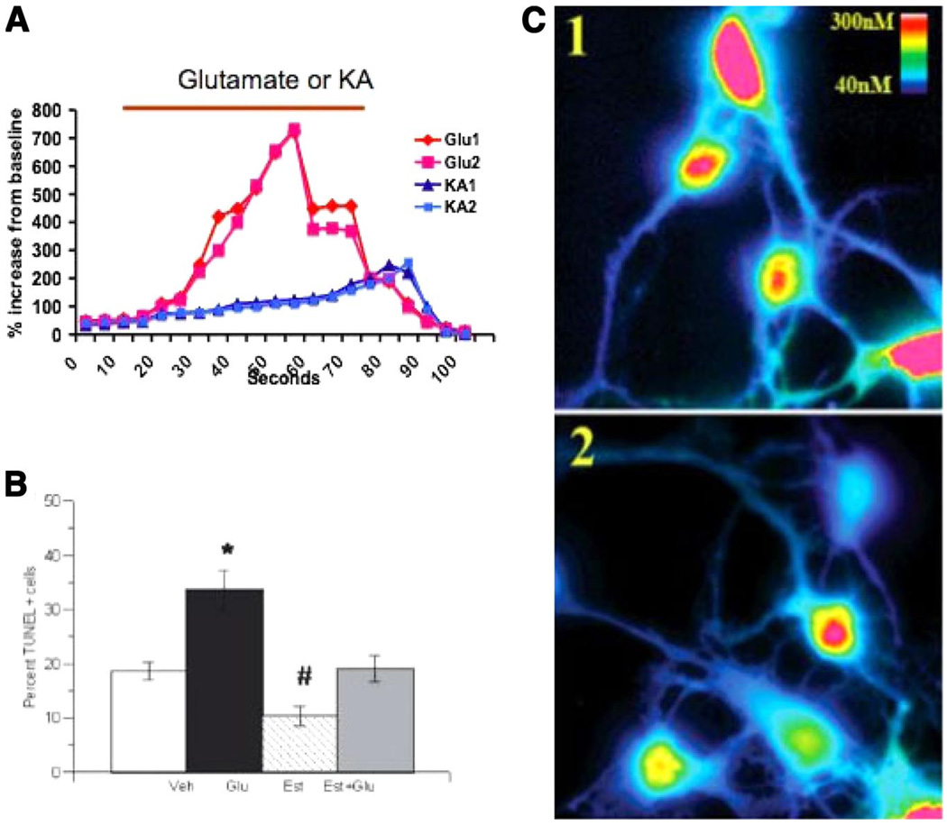
FIG 9.
Estradiol provides neuroprotection from glutamate excitotoxicity in immature brain. Excessive calcium influx following overactivation of glutamate receptors is the gateway to adult neuronal excitotoxicity. NMDA and AMPA receptors are primary routes for calcium influx into mature neurons. In immature hippocampal neurons, glutamate-mediated excitotoxicity is determined by the mGluR class of glutamate receptors and involves release of calcium from internal stores. A: neurons imaged with the calcium-sensitive dye fura 2-AM can be visualized over time to reveal the dynamics of calcium handling. Bath application of kanic acid (KA) versus glutamate (Glu) reflects the differential sources of calcium. KA activates AMPA, and then NMDA receptors and calcium influx into the cell with a slow upward slope that terminates gradually. In contrast, glutamate activates metabotropic glutamate receptors which release calcium from internal stores in a rapid surge that quickly depletes the reserve before the drug application is complete. B: treatment of cultured immature hippocampal neurons with glutamate significantly increases cell death, as indicated by TUNEL assay; pretreatment with estradiol completely prevents the glutamate-induced cell death and is neuroprotective against spontaneous cell death as well. C: pseudo-colored image of neurons being imaged for free calcium shows the control condition in panel 1, revealing a high level of calcium, whereas in panel 2, a decrease in the peak amplitude of the calcium is observed following pretreatment with estradiol. This reduction in the amplitude of the calcium transient correlates with an observed decrease in the amount of metabotropic glutamate receptor following estradiol treatment. [From Hilton et al. (93), copyright 2006 Wiley-Blackwell Publishing.]

