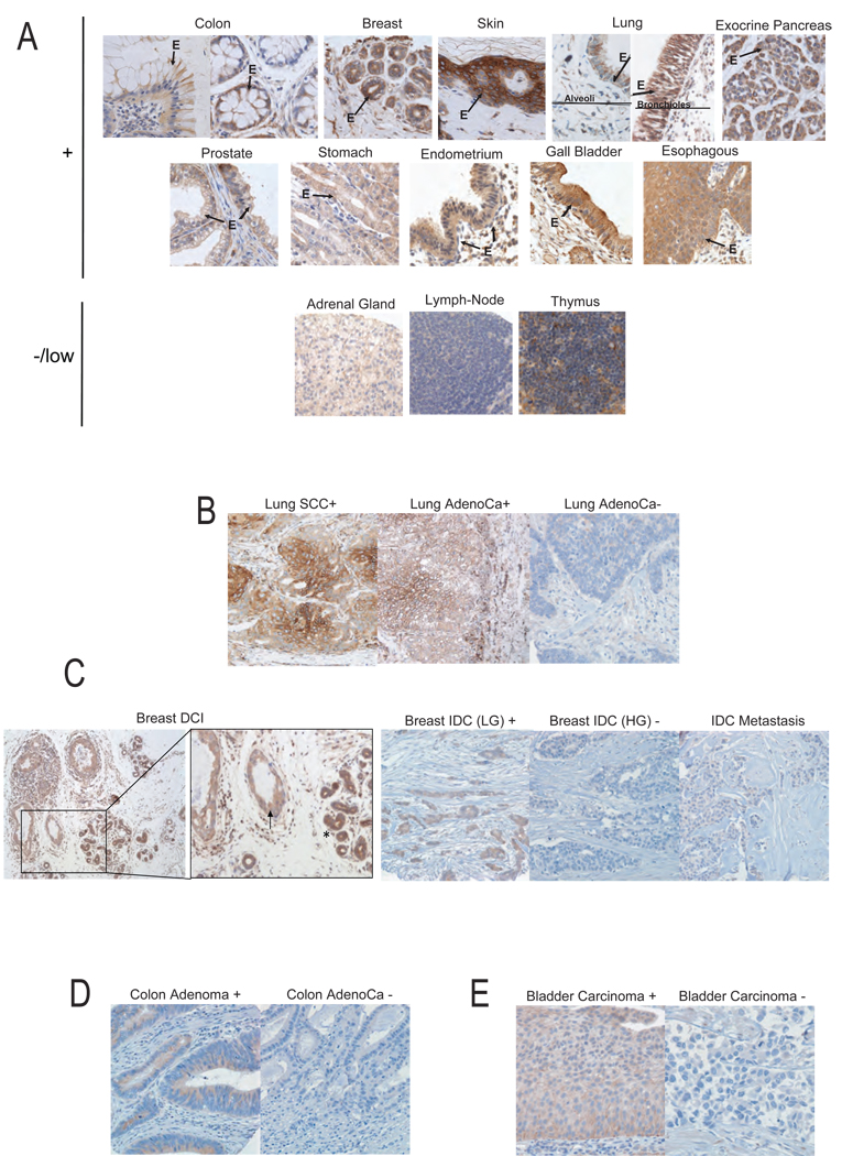
Figure 2. Cyfip1 expression in human normal and tumor tissues and its association with tumor progression.
Representative figures of Cyfip1 expression detected by immunohistochemistry (IH) in (A) normal tissues. The “E” and the arrows indicate the epithelium. (B) The majority of lung squamous cell carcinomas (SCC) and some adenocarcinomas (AdenoCa) showed a membranous-cytoplasmic expression of the protein (+), while most lung adenocarcinomas were found to have a negative phenotype (−). (C) Left panels show a ductal carcinoma in situ of the breast (DCI) (arrow) together with normal ducts (asterisk). Right panels show low grade (LG) invasive ductal breast carcinoma (IDC) exhibited intense and homogenous staining of Cyfip1, while most high grade (HG) IDC and bone metastatic adenocarcinoma lesions revealed a negative phenotype. (D) In colon tumor samples, high grade non-invasive adenomas maintain Cyfip1 while it is lost in a majority of invasive adenocarcinoma. (E) In bladder tumor samples, we observed that all low grade non-invasive superficial bladder carcinomas displayed a positive phenotype, while the majority of high grade invasive bladder carcinomas revealed undetectable Cyfip1 levels.

