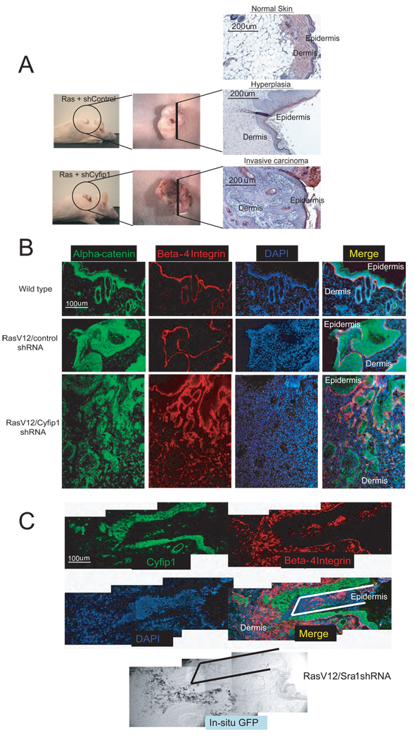
Figure 6. Knock-down of Cyfip1 promotes invasion in vivo.
Panel (A) illustrates macroscopically and microscopically the appearance of skin lesions produced in nude mice after transplantation of primary keratinocytes engineered to express oncogenic Ras or Ras plus Cyfip1 shRNA as compared with wild-type skin. (B) Markers of the epithelial compartment of the skin (β4-integrin and α-catenin) were used to study tissue architecture in sections from panel (A). (C) Immunofluorescence showing that invasive keratinocytes (β4-integrin positive) express the construct that contains the hairpin targeting Cyfip1 (positive for GFP mRNA in the in situ panel) and show low Cyfip1 expression. The lines (black, in merged panel and white, in in situ panel) are used as a reference.

