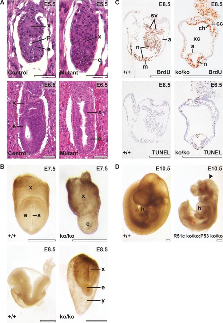
Figure 2. Rad51cko/ko mice die during early embryogenesis.
(A) Early postimplantation embryos sectioned with the deciduas and stained with hematoxylin and eosin. Top left, developmentally normal control E5.5 embrys reveals equally developed embryonic (e) and extraembryonic (x) tissues with a proamniotic cavity (p) clearly visible. Top right, mutant embryo demonstrates reduced embryonic tissues (e) without the cavity. At E6.5, development of embryonic tissues of the mutant embryo (bottom right) continues to lag behind compared with control embryos (Bottom left). (B) control (top left) and mutant (top right) embryos at E7.5. Primitive streak (s) is observed in control embryo. Morphology of control (bottom left) and mutant (bottom right) embryos at E8.5. x and e, as above; y, yolk sac. (C) Embryonic tissues in mutant embryo at E8.5 reveal active proliferation as evidenced by BrdU staining (top right) and increased apoptosis detected by TUNEL assay (bottom right) compared with control littermates (top and bottom left, respectively). n, neural folds; m, somites; a, amnion; sv, sinus venosus; xc, extraembryonic coelomic cavity; ch, prospective chorion; cc, ectoplacental cavity. Scale bar corresponds to 100 μm in A, and to 500 μm in B-D. (D) Partial rescue of the Rad51c-null phenotype on Trp53-deficient genetic background. Rad51cko/ko; Trp53ko/ko embryo at E10.5 (right) has almost normal morphology but smaller than a control Rad51cko/+; Trp53ko/+ littermate (left). Notice truncated caudal region and unclosed head folds in the mutant embryo (arrowhead). Mutant embryo shows apparently normal looking heart (h).

