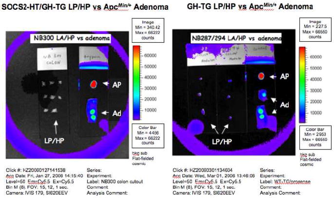
Figure 6. NIRF imaging of lymphoid polyps (LP) and hyperplastic polyps (HP).

NIRF images collected by a Xenogen IVIS system of dissected colonic lymphoid polyps (LP) and hyperplastic polyps (HP) from mouse NB300, a SOCS2-HT/GH-TG (Right) and mice NB287 and NB294, which were GH-TG (left). Note the isolated lymphoid and hyperplastic polyps had no NIRF signal. Activated Prosense™ 680 (AP) and adenomatous polyps (Ad) from Prosense™ 680 injected ApcMin/+ mouse colon imaged under identical conditions served as positive controls.
