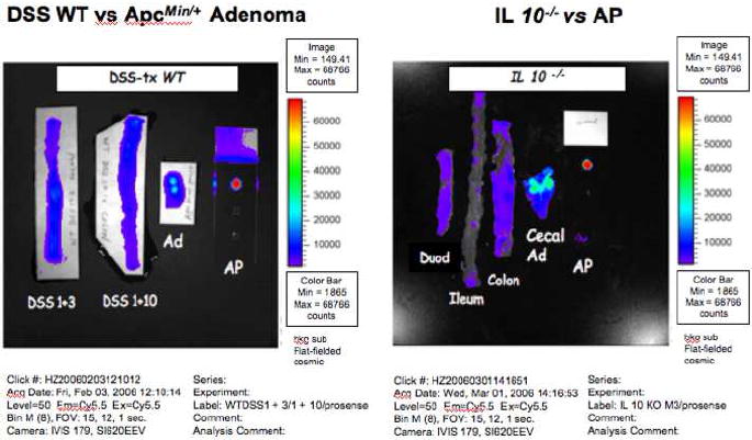
Figure 7. NIRF imaging of acute and chronic intestinal inflammation.

NIRF images captured by a Xenogen IVIS system of: Right = colon of DSS treated WT mice sacrificed 3 (DSS+3) or 10 days (DSS+10) after DSS treatment. Left = NIRF images of duodenum, ileum, colon and cecum of an IL-10-/- mouse. Note background signals except for what proved to be a cecal adenoma (Cecal Ad). The inflamed colon had no NIRF signals. Activated Prosense™ 680 (AP) served as positive control.
