
Figure 1. VSV infects Drosophila cells.
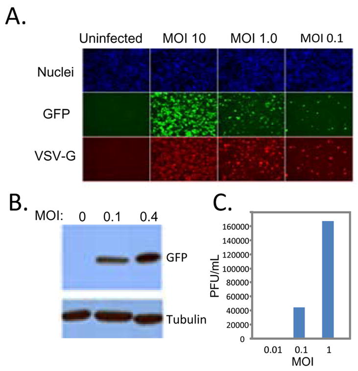
A. Schneider cells were infected with VSV-GFP at the indicated multiplicity of infection (MOI) for the 20 hours. Cells were processed for immunofluorescence and imaged using an automated microscope (ImagXpress Micro). Infected cells express GFP and the viral glycoprotein, VSV-G and are counterstained with Hoechst 33342 to observe nuclei. B. Viral antigen production at the indicated MOI at 24 hours post infection was measured by immunoblot against the virally produced antigen GFP or the cellular control tubulin. These data show a representative experiment; similar findings were made in at least three repetitions. C. Viral titers from cells infected with the indicated MOI of VSV at 24 hours post infection. These data show a representative experiment; similar findings were made in at least two repetitions.