Figure 4.
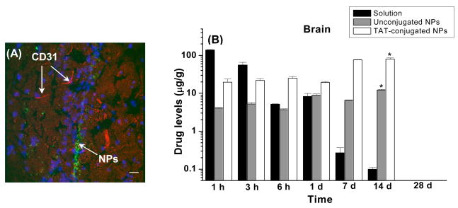
(A) Localization of 6-coumarin-loaded TAT-conjugated NPs in mice brains, 1 d after intravenous injection of NPs at a dose of 250 mg/kg. This dose of NPs is equivalent to the 45 mg/kg dose of ritonavir used in vivo study to determine brain uptake of the drug. Sections were observed using a confocal microscope equipped for fluorescence at 40X magnification under an oil immersion lens. Image demonstrates the localization of TAT-conjugated NPs within the brain ventricles. Magnification bar = 25 μm. Blue fluorescence is due to DAPI-staining of the nuclei, red fluorescence is due to CD-31 antibody-staining of the blood capillaries, and green fluorescence is due to the NP localization. Reproduced with permission from Rao et. al., Biomaterials 29:33 4429–38. (B): Brain levels of ritonavir in FVB/Ntac mice injected intravenously with either ritonavir solution or ritonavir-loaded unconjugated or TAT peptide-conjugated NPs at a drug dose of 45 mg/kg. Data are represented as mean ± s.e.m. (n = 4). * p < 0.05 compared between the TAT-conjugated NPs group with the unconjugated NPs and solution groups. Reproduced with permission from Rao et al. Biomaterials, (2008) 29:4429–38.

