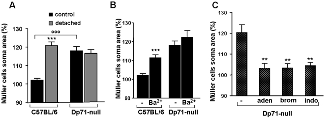
Figure 5. Osmotic swelling properties of Müller glial cells.
Müller cell somata were measured after 4 minutes perfusion of retinal slices with a hypotonic solution (60% of control osmolarity). Data were expressed in percentage of the values obtained before hypotonic challenge. A: Müller cells of control and detached retina from wt and Dp71-null mice were exposed to hypotonic challenge. In wt mice, only cells of detached retina swell. Note the swelling of Müller cells from control retina of Dp71-null mice. B: Müller cells of control retina from wt and Dp71-null mice were exposed to hypotonic challenge in the presence of barium chloride (Ba2+). Addition of Ba2+ induced a swelling of Müller cells of wt mice and had no effect on the soma area of Müller cells of Dp71-null mice. C: Addition of adenosine (aden); inhibitor of phospholipase A2, 4-bromophenacyl bromide (brom) and inhibitor of cyclooxygenase enzyme, indomethacin (indo) blocked the swelling of Müller cells of control retina from Dp71-null mice. Each bar represents value obtained in 12–34 cells; n = 3 mice for each group. Data are expressed as mean + SE. **p<0.01; ***p<0.001 significant differences versus control. °°°p<0.001 significant difference versus wt.

