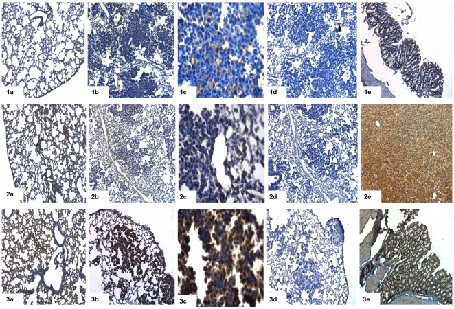
Figure 10. Immunohistochemical staining for adenocarcinoma (Cldn2, Fetub, Fst).
(a) Immunohistochemical staining of control lung tissue, (b) lung cancer at 10× magnification and (c) at 40× magnification (d) lung cancer in the presence of primary antibody, after preincubation with blocking peptide and (e) positive control. 1 = claudin 2 (Cldn2), 2 = fetuin beta (Fetub), 3 = follistatin (Fst).

