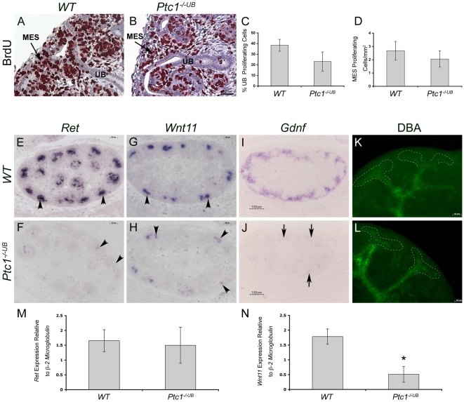
Figure 3. Ectopic HH signaling activity in the proximal epithelium impairs ureteric tip cell–specific gene expression.
(A–D) Analysis of cell proliferation in E13.5 kidney tissue using in situ BrdU incorporation assay. BrdU (brown color) is decreased in ureteric cells in Ptc1−/−UB kidney compared to WT. (C) Quantitative analysis of ureteric cell proliferation. Ureteric cell proliferation, quantitated as the percent of BrdU-positive ureteric cells, was decreased in Ptc1-deficient kidneys (p = 0.05). (D) Quantitative analysis of mesenchymal cell proliferation. Mesenchymal cell proliferation, quantitated as the number of BrdU positive cells per mm2 of renal tissue was comparable in WT and Ptc1−/−UB kidneys. ub = ureteric bud, mes = metanephric mesenchyme. (E–H) RNA in situ hybridization demonstrates expression of Ret and Wnt11 exclusively to the ureteric bud tips (arrowhead) in WT kidney tissue. In contrast, these mRNA transcripts are either absent or markedly reduced in Ptc1−/−UB kidney tissue (arrowhead). (I,J) RNA in situ hybridization demonstrates that Gdnf is markedly reduced in metanephric mesenchyme of Ptc1−/−UB kidneys (arrows). (K,L) DBA-lectin localizes predominantly to the ureteric stalk in WT kidneys and is excluded from the ureteric tips at E13.5. In Ptc1−/−UB kidneys DBA-lectin is observed throughout the ureteric tips and ureteric stalks. (M,N) Quantitative real-time PCR of isolated WT and Ptc1−/−UB ureteric buds at E11.5. (M) Ret expression is similar between WT and Ptc1−/−UB ureteric cells. (N) Wnt11 expression is significantly decreased in Ptc1−/−UB ureteric cells (p<0.05).

